Annals of Cardiology and Vascular Medicine
HOME /JOURNALS/Annals of Cardiology and Vascular Medicine- Case Report
- |
- Open Access
- |
- ISSN: 2639-4383
Ectopic Kidney: A Case Report on Sonographic Finding
- Solomon Demissie*;
- Department of Clinical Anatomy, Arba Minch University, Southwest, Ethiopia.
- Mulatie Atalay
- Department of Clinical Anatomy, Arba Minch University, Southwest, Ethiopia.

| Received | : | Jan 25, 2022 |
| Accepted | : | Feb 17, 2022 |
| Published Online | : | Feb 22, 2022 |
| Journal | : | Annals of Cardiology and Vascular Medicine |
| Publisher | : | MedDocs Publishers LLC |
| Online edition | : | http://meddocsonline.org |
Cite this article: Demissie S, Atalay M. Ectopic Kidney: A Case Report on Sonographic Finding. Ann Cardiol Vasc Med. 2022; 5(1): 1059.
Keywords: Ectopic kidney; Ultrasonography; Abaya clinic; Ethiopia.
Abstract
An ectopic kidney is the rare condition that occurs when the kidney fails to ascend from its embryologic position in the fetal pelvis to its final position in the renal fossa. This abnormal location leads to various disease conditions.
Case: A 13-year’s old male child came to clinics with a complaining of mass like distention around umbilical region and flank pain.
Result: Abdominally located kidney at right lumbar (2/3 of) and umbilical region (1/3 of) with poor corticomedullary differentiation on ultrasound examination.
Conclusion: The case has clinical importance for clinician, anatomist and researchers. Keywords: ectopic kidney, ultrasonography.
Introduction
The kidneys are a pair of bean-shaped organs located retroperitoneal on the posterior abdominal wall between the transverse processes of Thoracic vertebrae 12 and Lumbar vertebrae 3 [1]. Both kidneys upper poles are usually oriented slightly medially and posteriorly relative to the lower poles with slight difference in position where the right kidney is slightly inferior than the left kidney because of the liver [2].
The embryological development of the kidney occurs between the sixth to eighth weeks of life and the ascent of the kidney occurs during the ninth week [3].
There is a wide range of malformations resulting from disorders in the development process of kidneys. Among this ectopic kidney occurs when the kidney fails to ascend from its embryologic position in the fetal pelvis to its final position in the renal fossa [4].
In clinical practice, diagnosis of kidney is based on medical history, physical examination; laboratory test and radiological imaging using ultrasound, X-ray, Magnetic Resonance Imaging and Computed Tomography scan [5].
Case presentation
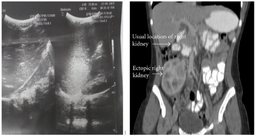
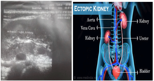
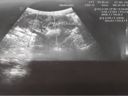
13 years old male child presented to outpatient department of Abaya medium clinics with complain of persistent urge to urinate, burning sensation when urinating and passing frequent, and small, red colored urine of one-week durations. The patient also complains infrequent abdominal distension, which persists for long period of time. The physical examination shows palpable abdominal tenderness umbilical region. The patient has no previous history of disease and it was first visit of clinics. The laboratory and ultrasound examinations were ordered for patient. The laboratory investigation of urine shows blood in the urine. The serum creatinine levels were within normal limits. The sonographic examination of the kidney shows that the left kidney is normal location, echo texture, well appreciated cortico- medullary differentiation and no dilation of pelvis and calyces of kidney (Figure 1). But on the right side the renal fossa shows no kidney. The scanning using linear probe on the umbilical region showed, well-defined kidney seen at right umbilical (1/3 of) and lumbar region (2/3 of) above iliac crest (Figure 2). The kidney is presented with poor corticomedullary differentiation (Figure 3). Based on finding the clinicians took previous familial history associated to ectopic kidney. But, the patient family respond that the case is unique even they didn’t hear such like cases. Finally, the physician treats the patient with antibiotics and advised to take fluids daily because the case is more susceptible for hydronephrosis, infection and nephrolithiasis. Follow up evaluation is also recommended for patient to comeback after one month for checkup.
Figure 1: sonographic images of a 13 years old patient with write empty renal fossa and normal location of left kidney in left renal fossa.
Figure 2: Sonographic images of a 13 years old patient with abdominally located right kidney on linear probe scanning.
Figure 3: Color Doppler sonographic image of right ectopic kidney located abdominally with poor corticomedullary differentiation.
Discussion
Congenital renal anomalies are among the 3rd most common birth defects [6]. The etiology behind ectopic kidney is the failures of the embryologic kidney migration from pelvis into the renal fossa in the 9th week [7]. The reason may be due to poor development of a kidney bud prompting the kidney to move to its usual position, genetic abnormalities, the mother’s exposure to an agent such as a drug or chemical during pregnancy and other teratogenic factors [8].
The location of ectopic kidney varies. It may be abdominal, lumbar or pelvic, based on its position in the retroperitoneum [9]. The abdominal kidney is very rare condition and more susceptible to infection due to its malrotations than pelveic kidney wich is similar to current study [10]. The reason of formation of infection anteriorly placed renal pelvis leading to impaired urinary drainage.
Declarations
Acknowledgments: The authors would like to acknowledge the Abaya Poly Clinic for allowing the study to be conducted, facilitate the study setting and for the unreserved offer of their medical equipment required for the data collection. We finally acknowledge the participant and the participant’s family for their cooperation.
Ethics Approval and consent to participate: The informed written consent was obtained from the participant’s parent because the age of the participant is below 18 years. After participant conformation, the permission letters were obtained from Abaya Poly Clinic to assess patient’s history from cards. Privacy and confidentiality of information were properly kept. All methods were in accordance with relevant guidelines and regulations.
Consent for publication: Written informed consent was obtained from the patient's legal guardian for publication of this case report and any accompanying images. A copy of the written consent is available for review by the Editor-in-Chief of this journal.
Availability of data and materials: All relevant data are included in the article. The dataset of this study is available from the corresponding authors upon reasonable request.
References
- Moore KL, Dalley AF. Clinically oriented anatomy: Wolters Kluwer India Pvt Ltd. 2018.
- El-Reshaid W, Abdul-Fattah H. Sonographic assessment of renal size in healthy adults. Medical principles and practice. 2014; 23: 432-436.
- Standring S. Gray’s anatomy E-Book: The anatomical basis of clinical practice: Elsevier Health Sciences. 2020.
- Oh KY, Holznagel DE, Ameli JR, Sohaey R. Prenatal diagnosis of renal developmental anomalies associated with an empty renal fossa. Ultrasound quarterly. 2010; 26: 233-240.
- Renard-Penna R, Marcy P, Lacout A, Thariat J. Imaging of the kidney. Bulletin du Cancer. 2012; 99: 251-262.
- Bakshi S. Incidentally detected pancake kidney: A case report. Journal of Medical Case Reports. 2020; 14: 1-6.
- Bhoil R, Sood D, Singh YP, Nimkar K, Shukla A. An ectopic pelvic kidney. Polish journal of radiology. 2015; 80: 425.
- Dutt SS. Ectopic Kidney/Renal Ectopia.
- Bauer SB. Anomalies of the kidney and ureteropelvic junction. Campbell’s urology. 1998: 1708-1755.
- Cinman NM, Okeke Z, Smith AD. Pelvic kidney: Associated diseases and treatment. Journal of endourology. 2007; 21: 836-842.
MedDocs Publishers
We always work towards offering the best to you. For any queries, please feel free to get in touch with us. Also you may post your valuable feedback after reading our journals, ebooks and after visiting our conferences.


