Annals of Cardiology and Vascular Medicine
HOME /JOURNALS/Annals of Cardiology and Vascular Medicine- Research Article
- |
- Open Access
Infective endocarditis in pediatric patients in a tertiary care hospital, Pakistan
- Arif Maqsood Ali;
- Department of Pathology and Blood Bank, Rawalpindi Institute of Cardiology, Pakistan
- Azhar Mehmood Kayani;
- Rawalpindi Institute of Cardiology, Pakistan
- Shazia Arif;
- Army Medical College, Rawalpindi, Pakistan
- Agha Babar Hussain
- Rawalpindi Institute of Cardiology, Pakistan

| Received | : | Aug 08, 2018 |
| Accepted | : | Sep 10, 2018 |
| Published Online | : | Sep 14, 2018 |
| Journal | : | Annals of Cardiology and Vascular Medicine |
| Publisher | : | MedDocs Publishers LLC |
| Online edition | : | http://meddocsonline.org |
Cite this article: Ali AM, Kayani AM, Arif S, Hussain AB. Infective endocarditis in pediatric patients in a tertiary care hospital, Pakistan. Ann Cardiol Vasc Med. 2018; 2: 1009.
Abstract
Introduction: Endocarditis is a rare disorder which is associated with significant morbidity & mortality world over especially in the developing countries. The etiology of disease has gradually shifted from Rheumatic heart disease to prosthetic & device associated endocarditis in the developed world. Very little is known about its etiology in Pakistan.
Objective: To find out the frequency of disease and organisms leading to endocarditis in children admitted in Rawalpindi Institute of Cardiology.
Methods: A retrospective cross sectional study was carried out in indoor pediatric patients at Rawalpindi Institute of Cardiology in Rawalpindi from Jan 1st till Oct 15th 2017. Out of 120 clinical suspected patients of endocarditis, only fifty patients fulfilled Modified Dukes Criteria for the diagnosis of endocarditis. Three sets of blood cultures were collected from the patients having fever before the start of antibiotics. Samples were incubated in Bact Alert 3D automated blood culture system (Biomereux France). All indicator positives were subcultured using standard microbiological methods. The blood cultures were followed daily & negative cultures were reported after 7 days of incubation. Echocardiographic findings were taken from patient’s clinical record. All the collected quantitative data were analyzed by SPSS 19.0.
Results: Endocarditis was diagnosed in 50/120 (41.6%) patients. There were 35 (70%) males & 15 (30%) females. The mean age of patients was 5.5(±1.7) years. The most common presenting complaints were fever, shortness of breath, chest discomfort & cyanotic spells. The common underlying disease associated with endocarditis was Congenital Heart Disease (CHD) (52%) followed by Rheumatic Heart Disease (RHD) (32%). Blood cultures were positive in 16 (32%) patients while in 34 (68%) patients, blood cultures were negative. Coagulase negative Staphylococcus was the most frequently isolated organism 5/16 (31.2%) in our studied patients.
Conclusion: Endocarditis is frequently diagnosed disease in our setup. CHD is the commonest underlying heart disease in our population. It is frequently complicated with infective endocarditis. The spectrum of disease has changed as Rheumatic heart disease is infrequently reported in our patients. Coagulase negative Staphylococcus is the most commonly isolated in blood cultures followed by Streptococcus viridians . Antibiotics used to treat children with endocarditis should be based on the local antibiogram & according to the current guidelines.
Keywords: Infective endocarditis; Congenital heart disease; Coagulase negative staphylococcus ; Blood culture
Introduction
Inspite of advances in medical, surgical and critical care interventions, Infective Endocarditis (IE) is still a life-threatening illness with a high morbidity and mortality. Endocarditis is a disease of the endocardium, the internal covering of the heart and heart valves [1]. It is caused when bacteria or other microorganisms get entry into the bloodstream and damage heart tissue. The patients with underlying cardiac disease, especially with defective or damaged heart valves are most prone to develop IE [2].
Common symptoms of endocarditis are intermittent fever with chills and body aches. Blood cultures and echocardiography are essential evidence to make final diagnosis. Specific intravenous antibiotics against microorganisms responsible for infection are the treatment of choice. Infective heart valves or an abscess may require surgery beside antibiotics. Endocarditis can lead to serious complications especially due to septic emboli in brain and other organs [3-5].
Positive blood culture and positive serology for Coxiellaburnetii are one of the major Modified Duke Criteria [6]. Common causative organisms of IE include Staphylococci, Streptococci, Enterococciand fastidious Gram negative coccobacilli. Mycobacteria, Rickettsia, Chlamydia and Fungi are infrequently reported. In recent studies, Staphylococci particularly Staphylococcus aureus has exceeded Streptococcus viridians as the most widely recognized etiological organism of infective endocarditis [7].
Studies from industrialized countries have shown a consistent improvement in survival of patients with IE over the past four decades, with mortality recently reported between 15%– 33%. A similar decrease in mortality is also seen in patients from the Indian subcontinent: 42% in 1970 and 23% in 2009 [8]. The prevalence of IE was estimated 0.34-0.64 cases per 100,000/ year, which reflects IE as an uncommon disease in children [9].
Very little is known about the etiology of endocarditis in Pakistan especially in children. Therefore, a study was carried out to find the frequency of disease & organisms leading to endocarditis in our setup.
Methods
A retrospective study was carried out in admitted pediatric patients with suspected IE at Rawalpindi Institute of Cardiology (RIC) Rawalpindi, Pakistan from Jan 1st till Oct 15th 2017. RIC is a 250 bed hospital tertiary care, centrally located cardiac hospital in twin capital city of Pakistan with a daily out patients of over 1000 adult patients and 35-50 pediatric patients. Out of 120 admitted clinically suspected patients, only fifty fulfilled modified Dukes criteria for the diagnosis of endocarditis [6]. Three sets of blood cultures were collected from the patients having fever before the start of antibiotics. Samples were in oculated in aerobic blood culture bottles and incubated in Bact Alert 3 D automated blood culture system (Biomereux France). All indicator positive blood cultures bottles were subcultured using standard microbiological methods to identify the organism and its antibiotic susceptibility using CLSI MS S 25. The blood cultures were followed daily & negative cultures were reported after 7 days of incubation. Serological testing was also carried out to confirm Brucella antibodies by latex agglutination test. Online software tools SPSS 19.0 were noted to calculate descriptive statistic and ANOVA. A P value (<0.05) considered statistically significant.
Results
A total of fifty patients diagnosed with endocarditis fulfilled Modified Duke’s criteria. There were 35 (70%) males & 15 (30%) females. The mean age of our studied patients was 5.5 (±1.7) years, ranging between 1.50 and 8.50 years. The most common presenting complaints were shortness of breath, chest discomfort & cyanotic spells.
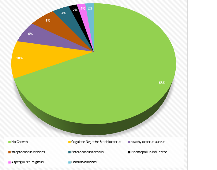
The most frequent predisposing disease associated with endocarditis was Congenital Heart Disease (CHD) 26(52%) followed by Rheumatic Heart Disease (RHD)16 (32%) (Table 1). Blood culture was positive in 16(32%) patients while the 34 (68%) patients were blood culture negative (Figure 1). Out of 16 positive blood cultures, the most frequently isolated organism was Coagulase negative Staphylococcus aureus 5/16 (31.2%) followed by Streptococcus viridians and Staphylococcus aureus 3/16 (18.7%) each. The organisms isolated in blood culture of patients with IE are shown in figure 1. The most common CHD seen were Ventricle Septal Defect (VSD) 9/26 (34.6%), Patent Ductus Arteriosus PDA 7/26 (27.0%),Tetrolgy of Fallot (TOF) 6/26 (23.1%) followed by others (Table 2). There is a significant correlation (p<0.05) found between disease type and positive blood cultures. No significant correlation was seen between positive blood cultures and age and gender (p>0.05).
Discussion
IE is a life threatening microbial infection of the endothelial surface of the heart. In spite of recent development in medical, surgical and critical care interventions, it is still associated with significant morbidity and mortality. In developed world a lot of research work is on going on endocarditis but very few studies have been reported from developing countries [2].
Previously, IE was seen in patients with valvular heart disease due to rheumatic disease. This was mainly seen in developing countries patients [8]. While in developed nations, IE was seen in patients with prior VSD, prosthetic valve, valvular stenosis, mitral valve prolapse, intravenous (IV) drug users and haemodialysis patients [10].
IE was considered a rare entity in children than adults. Over the last few decades the distribution of the disease has changed. Currently, CHD is the predominant cause of IE in children with overall decrease in RHD [9]. It is therefore important to carry out early microbiological diagnosis for rational use antibiotics therapy.The mean age of children with IE was 5.5(±1.7) years in our study. CHD was also found to be the most frequent predisposing disease leading to IE in our patients. The most common CHD seen were Ventricle Septal Defect (VSD) 9/26(34.6%), Patent Ductus Arteriosus PDA 7/26 (27.0%), Tetrolgy of Fallot (TOF) 6/26(23.1%) followed by others (Table 2). Our findings are similar to that reported by Ahmedi and Daryushi [11]. IE was more frequent in male patients and the dominant underlying causal risk factor was CHD. Dhjawan et al have reported similar results. There were 26 male compared to 11 female patients with IE in their study. Forty eight percent of their patients had underlying CHD. Six of their patients had extracardiac bacterial illness. Two of our patients did not have any cardiac predisposing disease. Both had a proceeding history of chest infection and pneumonia.
The prevailing distribution of IE in male population (70%) of our study was not considerably different with previous reported studies [9,11].
The most frequently isolated organism in blood cultures was Coagulase negative Staphylococcus 5/16(31.2%), Streptococcus viridians and Staphylococcus aureus 3/16(18.7%) each. Similar findings have been reported by Khan et al., who reported Coagulase negative Staphylococci in 35% cases, followed by Streptococci viridians 20% and Staphylococcus aureus in 17.5% patients [12]. Recent microbiological studies have reported that the rate of IE due to Staphylococcus species has increased while the incidence of IE by Streptococcus viridians has decreased [11,12]. Coagulase negative Staphylococcus was frequently isolated organism in our patients with IE. It has been primarily isolated in patients with prosthetic valves endocarditis particularly in those having fever, CHD, abscess and vegetations [12,13]. Our results are different from an African study from Tunisia where RHD is still the common predisposing disease of IE [14].
The HACEK organisms ( Haemophilus aphrophilus, Actinobacillus actinomycetemcomitans, Cardiobacterium hominis, Eikenella corrodens, and Kingella kingae ) were thought to be the most common agents involved in culture-negative endocarditis [15]. In a study from France more than 10% of all cases were found to be blood culture negative [16]. In our study 34 (68%) patients were blood culture negative. It is believed that these culture negative patients were possibly harboring HACEK organisms which were not identified. The most common reason for culture negative endocarditis might be due to frequent use of antibiotics by physicians to treat the condition empirically. Six patients were taking antibiotics among the culture positive endocarditis and 30 patients in culture negative endocarditis. Repeated blood culture before institution of antibiotics may increase the yield of culture positive results. For culture negative endocarditis serological and molecular methods can be used to find the possible HACEK like organisms.
IE is challenging and depends upon high index of suspicion. Use of Modified Dukes criteria is helpful in suspected cases of IE.
Conclusion
Endocarditis is a frequently diagnosed disease in our setup. CHD is commonest underlying heart disease that is often complicated with infective endocarditis. The spectrum of disease previously reported in our country has changed as few patients with IE had Rheumatic heart disease than CHD. Coagulase negative Staphylococcus is the most commonly isolated organism in blood cultures in patients with IE followed by Streptococcus viridians .
References
- Matta M, Mainardi JL. Endocardite infectieuse. Rev Prat. 2008; 58: 1029-35.
- Baddour LM, Wilson WR, Bayer AS, Fowler VG, Bolger AF, et al. Infective endocarditis: Diagnosis, antimicrobial therapy, and management of complications a statement for healthcare professionals from the Committee on Rheumatic Fever, Endocarditis, and Kawasaki Disease, Council on Cardiovascular Disease in the Young, and the Councils on Clinical Cardiology, Stroke, and Cardiovascular Surgery and Anesthesia, American Heart Association-Executive summary. Circulation. 2005; 111: 3167-84.
- Prendergast BD. Diagnostic criteria and problems in infective endocarditis. Heart. 2004; 90: 611-3.
- O’Gara PT. Infective endocarditis 2006: indications for surgery. Transactions of the American Clinical and Climatological Association. 2007; 118: 187-198.
- Pierce D, Calkins BC, Thornton K. Infectious endocarditis: diagnosis and treatment. American family physician. 2012; 85: 981- 986.
- Li SJ, Sexton DJ, Mick N, Nettles R, Fowler VG, et al. Proposed modifications to the Duke criteria for the diagnosis of infective endocarditis. Clin Infect Dis. 2000; 30: 633–638.
- Khan NU, Farman MT, Sial JA, Achakzai AS, Saghir T, et al. Changing trends of infective endocarditis. J Pak Med Assoc. 2010; 60: 24-7.
- Tariq M, Siddiqui BK, Jadoon A, Alam M, Khan SA, et al. Clinical profile and outcome of infective endocarditis at the Aga Khan University Hospital. International Journal of Collaborative Research on Internal Medicine and Public Health. 2009;1: 84.
- Knirsch W, Nadal D. Infective endocarditis in congenital heart disease. Eur J Pediatr. 2011; 170: 1111–27.
- Krul MM, Vonk AB, Cornel JH. Trends in incidence of infective endocarditis at the Medical Center of Alkmaar. Neth Heart J. 2015 Nov 1; 23: 548-54.
- Ahmadi A, Daryushi H. Infective endocarditis in children: A 5 year experience from Al-Zahra Hospital, Isfahan, Iran. Advanced Biomed Res. 2014; 3-228.
- Khan NU, Farman MT, Sial JA, Achakzai AS, Saghir T, et al. Changing trends of infective endocarditis. J Pak Med Assoc. 2010; 60: 24-27.
- Lalani T, Kanafani ZA, Chu VH, Moore L, Corey GR, et al. Prosthetic valve endocarditis due to Coagulase-negative staphylococci: Findings from the International Collaboration on Endocarditis Merged Database. Eur J Clin Microbiol Infect Dis. 2006; 25: 365–8.
- Jomaa W, Ali IB, Abid D, Ernez SH, Abid L, et al. Clinical features and prognosis of infective endocarditis in children: insights from a Tunisian multicentre registry. Archives of cardiovascular diseases. 2017; 110: 676-681.
- Sharara SL, Tayyar R, Kanafani ZA, Kanj SS. HACEK endocarditis: A review. Expert review of anti-infective therapy. 2016; 14: 539- 45.
- Hoen B, Selton-Suty C, Lacassin F, Etienne J, Briançon Set al. Infective endocarditis in patients with negative blood cultures: Analysis of 88 cases from a one-year nationwide survey in France. Clinical infectious diseases. 1995; 20: 501-6.
MedDocs Publishers
We always work towards offering the best to you. For any queries, please feel free to get in touch with us. Also you may post your valuable feedback after reading our journals, ebooks and after visiting our conferences.


