Annals of Cardiology and Vascular Medicine
HOME /JOURNALS/Annals of Cardiology and Vascular Medicine- Case Report
- |
- Open Access
- |
- ISSN: 2639-4383
One Case of Patient Took Warfarin for 10 Years after Biological Mitral Valve Biopsy
- Bailang Chen;
- Division of Cardiovascular surgery, The seventh affiliated hospital, Sun Yat-sen University, China
- Xianmian Zhuang;
- Department of Cardiovascular surgery, Fuwai Hospital, Chinese Academy of Medical Sciences, Shenzhen, China
- Minxin Wei*
- Department of Cardiovascular surgery, Fuwai Hospital, Chinese Academy of Medical Sciences, Shenzhen, China

| Received | : | Jun 05, 2020 |
| Accepted | : | Jul 03, 2020 |
| Published Online | : | Jul 07, 2020 |
| Journal | : | Annals of Cardiology and Vascular Medicine |
| Publisher | : | MedDocs Publishers LLC |
| Online edition | : | http://meddocsonline.org |
Cite this article: Chen B, Zhuang X, Wei M. One Case of Patient Took Warfarin for 10 Years after Biological Mitral Valve Biopsy. Ann Cardiol Vasc Med. 2020: 3(1); 1020.
Keywords: Chest; Breath; Mitral valve disease; Echocardiogram
Introduction
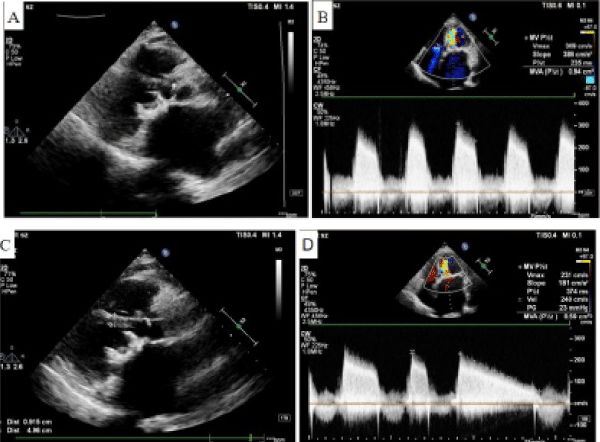
65-year-old female patient was admitted to the hospital due to chest tightness and shortness of breath. Patients underwent mitral valve replacement in 2010 due to “mitral valve disease”. The patient received the doctor’s advice for long-term, uninterrupted use of warfarin and went to the hospital regularly to review the echocardiogram and the valve was not abnormal. In September 2019, the patient listened to another doctor’s opinion and stopped Warfarin. The patient underwent echocardiography on November 20, 2019, suggesting that the bioprosthetic leaflets were thickened, calcified, and open limited. The maximum flow rate of the biological valve mouth is 3.1m/s, and
the peak pressure difference is 39 mmHg. (Figure A & B). Chest tightness, shortness of breath, both lower extremity edema occurred after the activity. The patient was treated in our hospital. After admission, he was given low molecular weight heparin calcium. After 0.4 ml q12h subcutaneous injection and diuretic treatment, the patient’s symptoms gradually eased. On December 5, 2019, the echocardiogram was reviewed, suggesting that the leaflets were still thickened but the opening was slightly restricted, The maximum flow rate of the biological valve mouth is 2.4 m/s, and the peak pressure difference is 23 mmHg. (Figure C & D). The patient’s valve condition was significantly improved. We did not give surgery to patients and recommended that patients take long-term warfarin. The patient is still in follow-up.
Figure 1: (A) Ultrasound manifestation of valve before anticoagulation therapy. (B) Echocardiography shows valve flow velocity is 3.1 m/s, the peak pressure difference is 39 mmHg. (C) Ultrasound manifestation of valve after anticoagulation therapy. (D) Echocardiography shows valve flow velocity is 2.4 m/s, the peak pressure difference is 23 mmHg.
Discussion
Due to its good hemodynamic characteristics and the advantages of no lifelong anticoagulation, the biological valve is widely used in clinical practice. With the advancement of technology, the life span of the biological valve is gradually prolonged, and the 12-year decay rate of the third-generation bioprosthesis reached (92±8)% [1]. But as time goes on, the valve gradually declines. The causes of biological valve decline are complex and diverse, and the current mechanism is still unclear. It is believed that bioprosthesis adhesion, abnormal metabolism of patients, early postoperative thrombosis, immune inflammatory reaction are the main causes of valve decline.
In this patient, there was no postoperative fever discomfort, and the rheumatoid factor was negative. The thrombus on the surface of the biological valve may have platelets and leukocyte deposition. The mitochondria of these cells are the sites where calcium crystals accumulate, which are easy to form calcification, and calcification can induce thrombus. The two complement each other, causing valve failure [2]. Infection of the biological valve often involves the valve leaflets, and the resulting neoplasm may block the valve orifice and cause stenosis. Due to infection, macrophages, white blood cells, platelets, etc are deposited on the leaflets, which also promotes leaflet stiffness and calcification. Degeneration of biological valves is mainly caused by calcification: How does this relate to antithrombotic therapy? One possible answer is leaflet thrombosis [3]. This hypothesis is supported by the pathophysiology of natural aortic stenosis, in which valvular lobular hemorrhage is associated with more rapid valvular calcification and disease progression [4].
There is still a lack of comprehensive research on whether anticoagulant therapy is effective in delaying the decay of biological valves. At present, for patients with bioprosthetic replacement, the thromboembolic complications are basically similar to those of mechanical valves within 4-6 months after surgery. Therefore, anticoagulant therapy should be performed within half a year after surgery. Occasional cause, this patient had long-term use of warfarin. After occasional discontinuation of warfarin, valve thrombosis occurred. After we re-administer anticoagulant therapy, the valve function is relieved. We believe that if the patient does not take warfarin anticoagulation for a long time, the valve may have decayed at an earlier time. From this perspective, lifelong anticoagulant therapy may have its benefits. At the same time, enhanced follow-up, early detection, early intervention. At present, although the biological valve does not require lifelong anticoagulation, the risk of lifelong anticoagulant therapy is not high for patients, and it is not particularly cumbersome. It does not need frequent detection after stabilization. If anticoagulation can delay the decline of biological valve, lifelong anticoagulation is a good choice. After discharge from the hospital, the symptoms of bacterial infection such as a cold may cause bioprosthetic thrombosis, and the patient is not taken seriously, thus accelerating valve decay.
Conclusion
In conclusion, Whether long-term anticoagulation is not needed for all patients with bioprosthetic replacement is questionable, and whether it is beneficial for long-term anticoagulation in such patients is worth studying. For patients with bioprosthetic replacement for many years, it is debatable whether patients with signs of decay in the valve need to add warfarin anticoagulant therapy at this time. It is difficult to accurately monitor changes in the patient's condition. For patients with bioprosthetic replacement, a more complete anticoagulant regimen may be used to better benefit patients, prolong valve use time, and reduce the occurrence of related complications in the future.
Declaration of patient consent
The authors certify that they have obtained all appropriate patient consent forms. In the form, the patient has given his consent for his clinical information to be reported in the journal. The patient understands that his name and initial will not be published and due efforts will be made to conceal his identity, but anonymity cannot be guaranteed.
References
- El Oakley R, Kleine P, Bach DS . Choice of Prosthetic Heart Valve in Today’s Practice. Circulation, 2008: 117: 253-256.
- Valente M, Bortolotti U, Arbustini E, Talenti E, Thiene G, et al. Glutaraldehyde-preserved porcine bioprosthesis: factors affecting performance as determined by pathologic studies. Chest. 1983; 83: 607-611.
- Doris M K, Dweck M R. Is bioprosthetic leaflet thrombosis a trigger to valve degeneration?. Heart.35 2018: heartjnl-2017-312861.
- Akahori H, Tsujino T, Naito Y, Matsumoto M, Lee-Kawabata M, et al. Intraleaflet haemorrhage is associated with rapid progression of degenerative aortic valve stenosis. European heart journal. 2011; 32: 888-896.
MedDocs Publishers
We always work towards offering the best to you. For any queries, please feel free to get in touch with us. Also you may post your valuable feedback after reading our journals, ebooks and after visiting our conferences.


