Annals of Cardiology and Vascular Medicine
HOME /JOURNALS/Annals of Cardiology and Vascular Medicine- Case Report
- |
- Open Access
Pigtail straightener left in ascending aorta
- Ravaux Justine Mafalda;
- Department of Cardiovascular Surgery, Grand Hospital De Charleroi, Gilly, Belgium
- Amond Laurent;
- Department of Cardiovascular Surgery, Grand Hospital De Charleroi, Gilly, Belgium
- Remy Philippe;
- Department of Cardiovascular Surgery, Grand Hospital De Charleroi, Gilly, Belgium
- Ghammad Kaoutar;
- Department of Cardiovascular Surgery, Grand Hospital De Charleroi, Gilly, Belgium
- Dasnoy Denis;
- Department of Cardiovascular Surgery, Grand Hospital De Charleroi, Gilly, Belgium
- Swaelens Charles
- Department of Cardiovascular Surgery, Grand Hospital De Charleroi, Gilly, Belgium

| Received | : | Aug 20, 2018 |
| Accepted | : | Oct 15, 2018 |
| Published Online | : | Oct 19, 2018 |
| Journal | : | Annals of Cardiology and Vascular Medicine |
| Publisher | : | MedDocs Publishers LLC |
| Online edition | : | http://meddocsonline.org |
Cite this article: Mafalda RJ, Amond L, Rémy P, Ghammad K, Dasnoy D, et al. Pigtail straightener left in ascending aorta. Ann Cardiol Vasc Med. 2018; 2: 1010.
Abstract
A case of retained Intra-Vascular Foreign Body (IVFB) after successful Thoracic Endovascular Aortic Treatment (TEVAR) for an isthmic penetrating atherosclerotic ulcer is reported. Thoracic pain on post-operative day one required angioscanner (CTA) investigations and showed a longilineal image from aortic valve to brachio-cephalic origin, confirmed by Trans-oesophageal Echo-Endoscopy (TEE). Endovascular re-intervention was performed with an Endovascular Snare. The object corresponded to the peel away straightener of the pigtail catheter used on the first intervention as angiography catheter. Patient was discharged on post-operative day 7 without complications.
Keywords: Endovascular treatment/therapy; latrogenic injury; Percutaneous; Safety; Pigtail straightener; Intravascular foreign bodies
Introduction
Intra-Vascular Foreign Bodies (IVFB) represent new increasing complications of endovascular intervention because of growing number of endovascular techniques, procedures and diversity of materials. As new complications imply new treatments, and consequently new clinical problems, highlights have to be made on such events to be as preventive as possible.
The first case of IVFB revealed a polyethylene catheter in the right atrium after autopsy [1]. Thomas et al [2] made the first publication about interventional non-invasive re-intervention for such a complication in 1964. Larger series [3] led to the development of many different materials to remedy the misplacement of the devices. Potential complications can occur in case of IVFB, such as vessel/cardiac perforation/lesions or thrombosis, groin hematomas, arrhythmias, haemoptysis, sepsis; sometimes leading to patient’s death [4-5]. Mortality rate related to IVFB is generally considered from 24% to 60% [6]. Risk factors were sorted as procedural-, patients-, safety variances- or equipment failure-related [7] and different causes were classified into three categories by Tateishi et al [8]: Inappropriate or inadequate techniques-, procedures regarding medical devices or devices or material problems- and others considerations. Success rate of treatment by endovascular approach is more than 90% [9]. Nowadays, the Snare is the preferred material for the retrieval of IVFB [10], making percutaneous endovascular treatment less invasive, simpler and safer confront to the surgical approach [11].
This case reports the problematic of IVFB illustrating an endovascular treatment for a peel away straightener of a pigtail catheter retained into the intravascular aortic sector after a TEVAR procedure. This work tries to set up some common attitudes in front of this emergent kind of complications.
Materials and methods
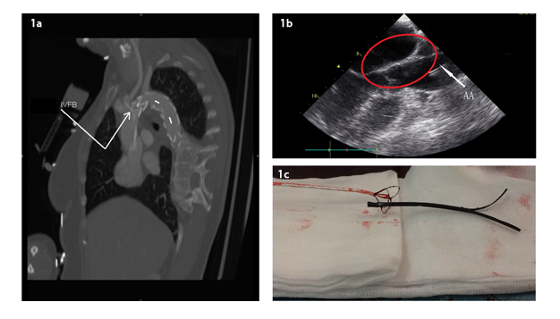
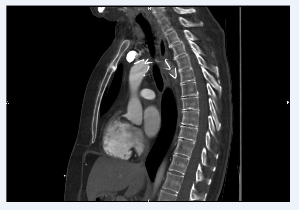
A 55-year-old female was admitted for dysarthia, sensation of cooling, numbness of the left arm and transient left face dyesthesia evolving for 1 hour. Relevant past medical history includes right aorto-femoral bypass currently occluded. CTA revealed an isthmic penetrating atherosclerotic ulcer with a focal dissection creating thrombosis on the left sub-clavian artery. A successful TEVAR with a Zenith TX2 Low-profil stent graft (Cook Medical, Bloomington, Ind.) was performed. After 24 hours, thoracic pain with inter-scapular irradiation required a new CTA, showing a hyper-intense and longilineal image from the aortic valve to the brachio-cephalic trunk origin (Figure 1a), confirmed by TEE demonstrating a lumen catheter (Figure 1b). The image indicated an IVFB but the nature of the IVFB was not clear at this moment. A re-intervention in order to remove this IVFB by the same previous endovascular approach using an endovascular Snare System (EN Snare, Merit Medical Systems, Inc., South Jordan UTl) was performed opening up the peel away straightener (Figure 1c) of the pigtail catheter (Cook Medical, Bloomington, Ind.) used for angiography. After 24 hours post-operative monitoring in intensive care unit for control, patient was discharged on the post-operative day 7. There were no adverse events during the recovery period. CTA control at six months showed a good position of the endoprosthesis without residual dissection or endoleak (Figure 2).
Figure 1: a. Post-operative day one thoraco-abdominal CTA showing a hyperintense and longinileal image from the aortic valve to brachio-cephalic origin (IVFB). b. Presence of IVFB confirmed by TEE (red circle) into the aortic arch (AA). c. Peel away straightener of the pigtail corresponding to the IVFB once removed.
Results and discussion
IVFB is a growing emergent complication in the area of endovascular surgery. In 2002, the National Quality Forum in the United States listed serious reportable and preventable events including unintentionally retained vascular devices in the Never Events [4]. Nevertheless, complexity and heterogeneity of such events create lack of prevention measures, diagnostic methods and general guidelines.
In this case, the aorto-femoral bypass presently occluded on the contralateral limb constrained us to a unique vascular access for the initial TEVAR procedure. This access imposed the use of the Flexor Sheath from the initial Stent Graft to obtain a final angiography by replacement of the pigtail catheter. The most likely hypothesis is that the peel away straightener of the pigtail catheter remained into the sheath and was consequently pushed up to the ascendant aorta during this final manipulation.
Making the diagnosis was difficult. The most common IVFB is a fragment of central venous catheter [6], but CTA didn’t confirm this hypothesis and showed no retrograde extension of the initial dissection. TEE was performed because of its high ability in diagnosing diseases of the thoracic aorta and for excluding structural defects/imperfections of the endoprosthesis patency [12].
Making the decision for a re-intervention is always challenging because of the necessity to balance carefully risks and benefits for the patient [13]. The thoracic pain of the patient and the inter-scapular irradiation with an image of IVFB on the CTA and the TEE led us to the re-intervention. The proximity with the aortic valve made the procedure tricky. A smaller Flexor introducer (Cook Medical, Bloomington, Ind.) was used in the left femoral artery to reach the aortic segment between aortic valve and brachio-cephalic origin with the Endovascular Snare Trifolia. After numerous attempts to capture the IVFB into the 3 interlaced loops, distally overtaking of the IVFB with the snare was the only way to retrieve this one, risking of making valve injuries by opening the snare. Moreover, distally slipping the IVFB gradually could hurt the aortic wall. Success was achieved and the integral peel away straightener was removed. This technique can be assimilated to the “Lateral Grasp Technic” described by J.B. Woodhouse in 2013: Distal deployment of the snare to the lesion following opening to remove the IVFB after using a rigid guide wire on the other side of the IVFB passing through the snare loop to pinch it and then to remove together guide wire and snare [13].
According to our experience, multidisciplinary approach with meticulous quality control is mandatory [12-14] as well as collaboration between all clinicians. Per-operative communication is crucial, even in emergency conditions. A material-checklist should be made after each endovascular procedure. Sheath introducers (whatever the label) and pigtail catheters are commonly used for endovascular procedures. We have to keep in mind that a part of the material can be let in the intravascular compartment and make sure that no piece of the device is lacking at the end of the intervention. That is why some ascertainment should be made during the procedure as we tried to list in table 1.
Table 1: Material-check-list of devices and implants usually in endovascular procedure to check before leaving the operating room and per-operatively. This table does not substitute the usual nursing/medical-check-list used for every kind of intervention (presentation of the patient, actors).
Conclusions
To our knowledge, this is the first case relating such an IVFB, illustrating the growing heterogeneity of IVFB and the increasing diverse management of the problem. Some general guidelines have to be published to reduce incidence and approximation in this area.
References
- Turner DD, Sommers SC. Accidental passage of a polyethylene catheter from cubital vein to right atrium; report of a fatal case. N Engl J Med. 1954; 251: 744-745.
- Thomas J, Sinclair-Smith B, Bloomfield D, DAVACHI A. A. Nonsurgical retrieval of a broken segment of steel spring guide from the right atrium and inferior vena cava. Circulation. 1964; 30: 106-108.
- Dondelinger RF, Lepoutre B, Kurdziel JC. Percutaneous Vascular Foreign Body Retrieval: Experience of an 11-year period. Eur J Radiol. 1991; 12: 4-10.
- Whang G, Lekht I, Krane R, Peters G, Palmer SL, et al. Unintentionally retained vascular devices: Improving recognition and removal. Diagn Interv Radio. 2017; 23: 238-244.
- Schechter MA, OBrien PJ, Cox MW. Retrieval of iatrogenic intravascular foreign bodies. J Vasc Surg. 2013; 51: 276-281.
- Halkati P, Patted S. Modi R, Tasgaonkar R. Retrieval of devices – Percutaneous tecniques. Int J Sci Res (Ahmedabad). 2015; 5.
- Moffatt-Bruce SD, Ellison EC, Anderson HL, Chan L, Balija TM, et al. Intravascular retained surgical items: A multicenter study of risk factors. J Surg Res. 2012; 178; 519-523.
- Tateishi M, Tomizawa Y. Intravascular foreign bodies: Danger of unretrieved fragmented medical devices. J Artif Organs. 2009; 12; 80-89.
- Rodt T, Von Falck C, Wilhelmi M, et al. Interventional removal of intravascular foreign bodies. Poster Session presented at : European Congress of Radiology ; 2012; 01-05.
- Carroll MI, Ahanchi SS, Kim JH, Panneton JM. Endovascular foreign body retrieval. J Vasc Surg; 2013; 57: 459-463.
- Motta Leal Filho JM, Carnevale FC, Nasser F, Santos AC, Sousa Junior Wde O, et al. Endovascular techniques and procedures, methods for removal of intravascular foreign bodies. Rev Bras Cir Cardiovasc. 2010; 25: 202–208.
- Fisher CH, Campos FO, Palma JH, Rodrigues Alves CM, Marcondes Sousa JA, et al. Use of transoesophageal echocardiography during implantation of aortic endoprothesis (stent): Initial experience. Arq Bras Cardiol ; 2001; 77: 5-8.
- Woodhouse JB, Uberoi R. Techniques for intravascular foreign body retrieval. Cardiovasc Intervent Radiol. 2013; 36: 888-897.
- Cho S-R, Tisnado J, Beachley MC, Vines FS, Alford WL, et al. Percutaneous unknotting of intravascular catheters and retrieval of catheter fragments. AJR. 1983; 141: 397-440.
MedDocs Publishers
We always work towards offering the best to you. For any queries, please feel free to get in touch with us. Also you may post your valuable feedback after reading our journals, ebooks and after visiting our conferences.


