Annals of Cardiology and Vascular Medicine
HOME /JOURNALS/Annals of Cardiology and Vascular Medicine- Case Report
- |
- Open Access
- |
- ISSN: 2639-4383
Rupture of dissected ascending aorta in redo patients also happens and has manifestations like pulmonary embolism
- Hadi abdulsalam Abo Aljadayel*;
- Master cardiac surgery, Syrian and Arab Board cardiac surgery, Syria
- Alireza Yaghoubi;
- Iranian board in cardiac surgery
- Sadegh Zargar Nataj;
- Iranian board in cardiac surgery
- Kourosh Trigarfakheri
- Iranian board of anesthesia

| Received | : | Mar 10, 2020 |
| Accepted | : | Apr 17, 2020 |
| Published Online | : | Apr 21, 2020 |
| Journal | : | Annals of Cardiology and Vascular Medicine |
| Publisher | : | MedDocs Publishers LLC |
| Online edition | : | http://meddocsonline.org |
Cite this article: Abo Aljadayel HA, Yaghoubi A, Nataj SZ, Trigarfakheri K. rupture of dissected ascending aorta in redo patients also happens and has manifestations like pulmonary embolism. Ann Cardiol Vasc Med. 2020: 3(1); 1015.
Keywords: Aortic surgery; Aortic dissection; Pulmonary compression; Previous cardiac surgery
Abstract
Most physicians believe that ascending aortic dissection in Redo cases never rupture because of adhesion and its relatively not emergent case but it seems not so. We report a case of 65-year-old woman who underwent CABG 5 years ago and came to emergency room with signs of dyspnea, tachypnea, tachycardia, chest pain. Because of adhesion Rupture of redo cases happens in posterior wall of aorta and compress the pulmonary artery to cause uncommon pulmonary embolism like signs which had never reported before and that clinicians should be aware of.
Introduction
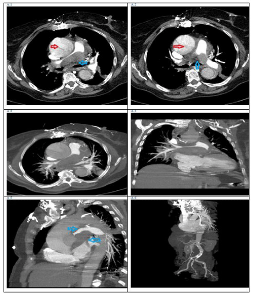
A 65-year-old woman brought to emergency of our hospital with fatigue, chest pain, dyspnea, tachypnea, tachycardia, and discomfort for 5hours; her symptoms were progressively worse during any movement then also at rest. She had a history of hypertension for 10 years with blood pressure of 160/90 mm Hg, and Coronary artery bypass graft 5 years before. Her arterial blood gases referred to respiratory acidosis. The patient’s temperature was 36.8C, pulse was 108beats/min, respiratory rate was 36 breaths/min, and blood pressure was 95/50 mm Hg. Slightly lower breath sounds were heard in the right lung. Two hours after hospitalization, tachypnea and dyspnea increased and the patient intubated electively in the CCU unit. The lactate was high and the D.dimer test was positive and high, also the ABG refers to acidosis. then the chest computed tomography angiography showed that the ascending aorta and aortic arch have intimal dissection flap and there was mass in the space between ascending aorta and pulmonary trunk which compress the main pulmonary trunk and right pulmonary artery. Figure-1A [1-6].
Management
According to the patient’s medical history, imaging manifestations, and acute reduction of lung function, rupture of acute thoracic aortic dissection with retro aortic hematoma and the compress of the right pulmonary artery was diagnosed. The patient’s family, concerned with the patient’s emergent situation and high risk of surgery and they accept the surgery. The patient transferred to operation room and operated.
In surgical ICU the patient was stable and in the third day extubated
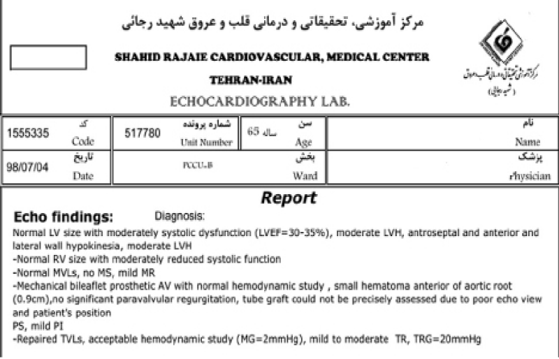
Trans thoracic echo was in normal and acceptable surgical results Figure (2) in the 4th day the patient developed respiratory distress and chest X-Ray and Ct scan revealed right lower lobe collapse and atelectasis.
Sonograghy was done to see the diaphragm movement which reveals normal movements in tow side of the diaphragm the patient re-intubated for another 72 hours then extubated again with normal chest X-ray and normal blood gases.
PORTABLE SONOGRAPHY OF DIAPHRAGM
By trans thoracic approach with 3.5 MHZ probe;
Both hemi diaphragms have normal movement.
there is no evidence of diaphragmatic paresis.
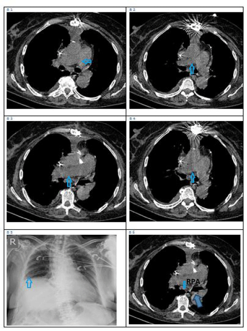
Two days later transferred to ward and After 2 weeks of ongoing medical and physiotherapy she discharged to home with normal parameters. The patient’s general situation was improved. At 2 months follow-up, the patient had no chest discomfort. Chest computed tomography showed normal exists and the pulmonary artery was open. Figure 3B [1-6].
Figure 1: CT angiography for the patient reveals the Ascending aorta dissection (red arrow) and the hematoma witch compress the pulmonary artery (blue arrow).
Figure 2: Trans thoracic post operation echocardiography report reveal the acceptable correction for ascending Aorta dissection and tricuspid valve repair without any injury for the L.V function.
Figure 3: post operation CT scan (B1,2,3,4,6)reveals that the compression of pulmonary artery removed by correcting the aortic dissection, and CXR (B5) in which we can see collapse in the right plural field.
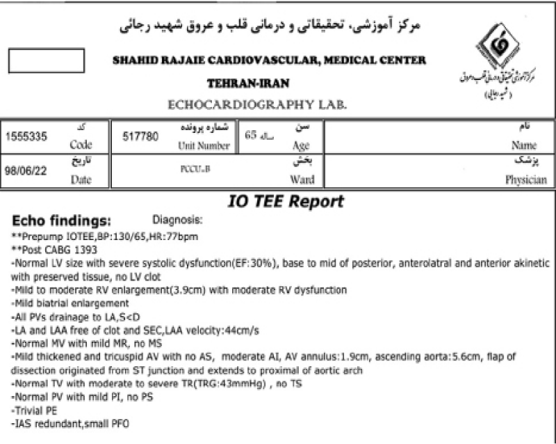
Figure 4: Trans esophageal echocardiography intra-op pre pump reveals the poor L.V dysfunction before operation and ascending Aorta dissection with severe T.V regurgitation.
Discussion
Most physicians believe that ascending aortic dissection in Redo patients never rupture because of adhesion and because of that mostly these patients operated as urgent list or as soon as possible patient more than emergency cases [1,2]. The rupture of aortic dissection in previously operated patients (redo cases) and compressing the pulmonary artery didn’t reported before but some articles speak about fistulas between aorta and pulmonary artery or right atrium or rarely pericardium in chronic dissection or late managed cases in patients with previous cardiac surgery[1,2].
Many studies talk about conservative nonsurgical treatment for patients who had dissection after previous cardiac surgery [1,2,4].
In this case, because of previous CABG a lot of adherent around the aorta helped to prevent wide rupture and the rupture was limited to retro aortic free space. Surgical intervention was performed promptly as in primary aortic dissection type A [2,4,5].
The exact mechanism involved in the post operation pulmonary collapse is not clear as the sonography revealed normal diaphragm movements and there was no foreign body in the bronchus endoscopy and no signs of infection. Clearly we don’t know if there is relationship between this and the compression over the right pulmonary artery before operation.
Operation technique
Intra operation Trans esophageal echocardiography revealed ascending aorta dissection with severe tricuspid valve regurgitation and low Ejection fraction and moderate right ventricle dysfunction Figure 4.
with femoral canulation the sternotomy was done on Bypass ascending aorta was dilated, superior vena cava was canulated and used tapping around SVC, IVC , during hypothermic time the tricuspid valve was explored on beating heart and repaired up to DeVega technique.
Retro grade cannula inserted directly to the coronary sinus for giving cardioplegic solution. The ascending aorta was closed and opened to see very big ruptured in the ascending aorta intima and the posterior wall of aorta was ruptured and a 10*10 cm hematoma was seen compress the pulmonary artery. After giving cardioplegia directly in coronary ostiums retrograde. The hematoma which consists of new clots removed. The previous two vein grafts were isolated as two small bottoms. Aortic valve replaced with mechanical St-jude 23 aortic valve then the ascending aorta replaced supracoronry with Dacron tube graft 30mm.
The pulmonary artery was opened and check for clots but it was clean so it’s closed.
At 20c degree total circulatory arrest was done aortic clamp removed and tow cannulas was advanced to the innominate and left carotid arteries to perfuse the brain with 600CC of blood per minutes.
Hemi arch replacement was done. All suture lines were supported with felt strep and new media technique was achieved to close false lumen.
Conclusion
Redo ascending aortic dissection is emergency operation and must operate promptly like primary cases. Because of adhesion Rupture of redo cases happens in posterior wall and compress the pulmonary artery to cause uncommon pulmonary embolism like signs that clinicians should be aware of.
References
- Norton EL, Rosati CM, Kim KM, Wu X, Patel HJ, et al. Is Previous Cardiac Surgery A Risk Factor for Open Repair of Acute Type a Aortic Dissection? The Journal of Thoracic and Cardiovascular Surgery. 2019.
- Estrera AL, Miller CC, Kaneko T, Lee TY, Walkes JC, et al. Out comes of acute type A aortic dissection after previous cardiac surgery. AnnThorac Surg. 2010; 89: 1467-1474.
- Klodell CT, Karimi A, Beaver TM, Hess PJ, Martin TD. Outcomes for acute type A aortic dissection: effects of previous cardiac surgery. Ann Thorac Surg. 2012; 93: 1206-1212.
- Elizabeth L, Norton MS, Rosati CM, Kim KM, Wu X, et al, Mich; Is previous cardiac surgery a risk factor for open repair of acute type A aortic dissection? The Journal of Thoracic and Cardiovascular Surgery, 10.1016 jtcvs.2019.07.093
- Hostiuc S, Dermengiu D, Ceaus UM, Capatina ACO, Lacramioara Luca , Mihaela Hostiuc c Sudden death due to dissection of the thoracic aorta associated with dissection and rupture of the pulmonary artery: Report of two cases, S. Hostiuc et al. / Forensic Science International. 2014; 236: e9–e13.
- Hirose H, Svensson LG, Lytle BW, Blackstone EH, Rajeswaran J, et al. Aortic Dissection After Previous Cardiovascular Surgery, Ann Thorac Surg. 2004; 78: 2099-2105.
MedDocs Publishers
We always work towards offering the best to you. For any queries, please feel free to get in touch with us. Also you may post your valuable feedback after reading our journals, ebooks and after visiting our conferences.


