
IMPACT FACTOR: 1.8
Short Name: J Radiol Med Imaging
ISSN: 2637-885X
Publisher: MedDocs Publishers LLC
Breast cancer is the most commonly diagnosed cancer and is the leading cause of death in women globally with an estimated 2.3 million new cases [1]. According to the cancer statistics, the incidence rate of breast cancer increased slightly by 0.3% per year over the recent five- year (2012-2016) [2].
The skull base angle, a critical anatomical and clinical parameter, plays a pivotal role in various medical disciplines, including neurology, otolaryngology, and maxillofacial surgery. Understanding the skull base angle involves examining the intricate anatomical relationships within the cranial base.
CTDI measures the radiation dose for each individual slice, DLP reflects the cumulative dose across the entire scanned region. Together, these metrics provide a standardized framework for assessing radiation levels, facilitating comparison across different scans and institutions, and ensuring adherence to safety standards.
Intussusception is a rare but potentially life threatening condition in children. Colocolic intussusception is extremely uncommon in pediatric patients and is usually associated with a pathological lead point.
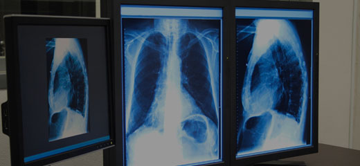
Human echinococcosis primarily affects the lungs, with Multiple Pulmonary Echinococcosis (MPE) occurring in 12% of cases. A rare manifestation, "cannonball-like" opacities, arises from bronchogenic dissemination after cyst rupture. Chest X-ray shows multiple, well-defined, nodular opacities
We always work towards offering the best to you. For any queries, please feel free to get in touch with us. Also you may post your valuable feedback after reading our journals, ebooks and after visiting our conferences.

